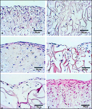
Histological micrographs of collagen–GAG matrices seeded with articular chondrocytes (a–d) and meniscus cells (e–f) after 21 days in culture. Scale bars are 50 µm. Read more about this figure in Zaleskas, J. M., B. Kinner, et al. "Growth Factor Regulation of Smooth Muscle Actin Expression and Contraction of Human Articular Chondrocytes and Meniscal Cells in a Collagen–GAG Matrix." Experimental Cell Research 270, no. 1 (2001): 21–31. (Courtesy of Elsevier, Inc., http://www.sciencedirect.com. Used with permission.)
Instructor(s)
Prof. Myron Spector
Prof. Ioannis Yannas
MIT Course Number
2.785J / 3.97J / 20.411J / HST.523J
As Taught In
Fall 2014
Level
Graduate
Course Description
Course Features
Course Description
Mechanical forces play a decisive role during development of tissues and organs, during remodeling following injury as well as in normal function. A stress field influences cell function primarily through deformation of the extracellular matrix to which cells are attached. Deformed cells express different biosynthetic activity relative to undeformed cells. The unit cell process paradigm combined with topics in connective tissue mechanics form the basis for discussions of several topics from cell biology, physiology, and medicine.


