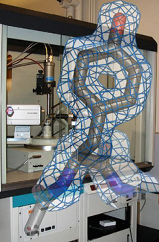
Molecular model of the amino acid tyrosine with experimental electron density in front of an X-ray diffractometer at MIT. The tyrosine is part of the crystal structure of phosphoglycerate mutase from M. tuberculosis. See Mueller, P., et al. Acta Cryst D61 (2005): 309-315. (Figure and photograph by Dr. Peter Mueller.)
Instructor(s)
Dr. Peter Mueller
MIT Course Number
5.069
As Taught In
Spring 2010
Level
Graduate
Course Description
Course Features
Course Description
This course covers the following topics: X-ray diffraction: symmetry, space groups, geometry of diffraction, structure factors, phase problem, direct methods, Patterson methods, electron density maps, structure refinement, how to grow good crystals, powder methods, limits of X-ray diffraction methods, and structure data bases.
Other Versions
Other OCW Versions
Archived versions: ![]()


